Our Science
NKT SCIENCE:
Target the Immune Response Earlier in the Inflammatory Cascade to Interrupt Disease Progression
Natural killer T (NKT) cells are innate-like T cells that share properties of both NK cells and T lymphocytes. NKT cells are pre-loaded with cytokine message and respond quickly in immune responses, but also help to maintain and propagate chronic long term immune responses. They interact with and influence the activity of other cell types and are a functional link between the innate and adaptive immune systems. iNKT are pro-inflammatory effector T cells that are pathogenic in many chronic fibrotic diseases such as idiopathic pulmonary fibrosis, lupus nephritis and NASH. dNKT cells are anti-inflammatory regulatory T cells that can reset unwanted immune responses that contribute to many autoimmune disorders such as systemic lupus erythematosus, multiple sclerosis and rheumatoid arthritis. GRI has a robust portfolio of patented drug candidates that regulate NKT cells.
NKT Cells for Immune Regulation
A novel immune mechanism to regulate the adaptive-innate immune axis and reset dysregulated immune responses.
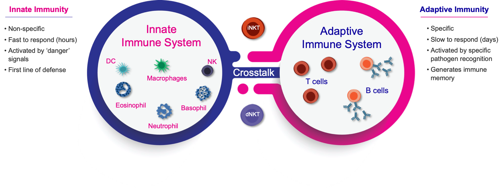
Figure 1. NKT cells are innate-like T cells that bridge the adaptive and innate immune systems.
NKT cells are innate-like adaptive T cells that influence the activity of other immune cells and help mediate the crosstalk between the adaptive and innate immune systems. iNKT cells behave as pro-inflammatory effector T cells in chronic inflammatory conditions and dNKT cells as anti-inflammatory regulatory T cells.
Regulating NKT Cells is a Selective Approach to Immunomodulation via Resetting the Immune Response
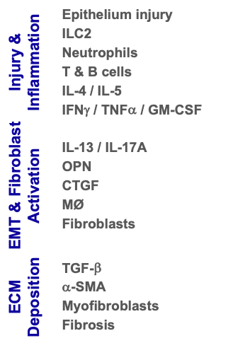
Targeting iNKT Cells Upstream of Key Fibrotic Mediators Provides Significant Benefits
iNKT play a key role in propagating the inflammatory / fibrotic cascade and downregulating them provides potential for downstream benefits, including immune resolution and homeostasis.


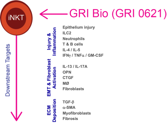
iNKT Cells play a Key Role in Fibrosis Progression in Patients
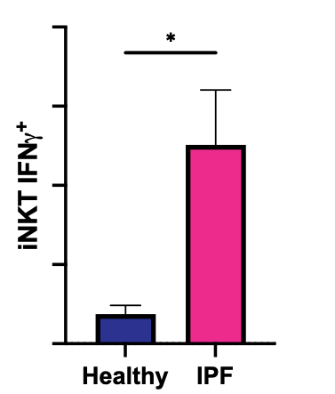
IPF
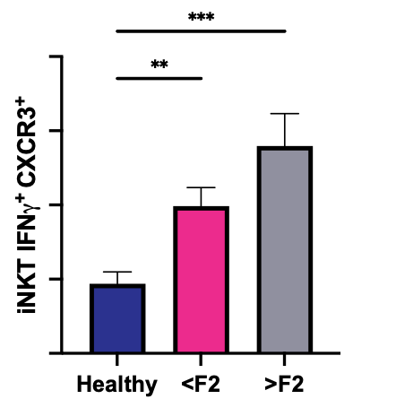
MASH
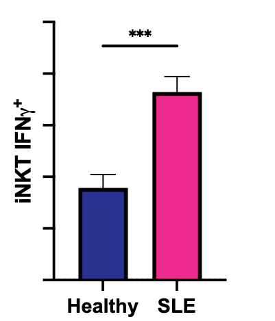
Lupus
Activated iNKT cells are increased in PBMC samples from IPF, NASH and SLE patients compared to healthy subjects.
Activated iNKT cells are increased in PBMC samples from IPF, NASH, and SLE patients compared to healthy volunteers or controls. Notably, activated iNKT cells increase in NASH patients with worsening fibrosis and worsening NAS scores (data not shown).
iNKT Cell Deficiency Reduces Expression of Key Fibrogenic Genes in a Model of Hepatic Fibrosis
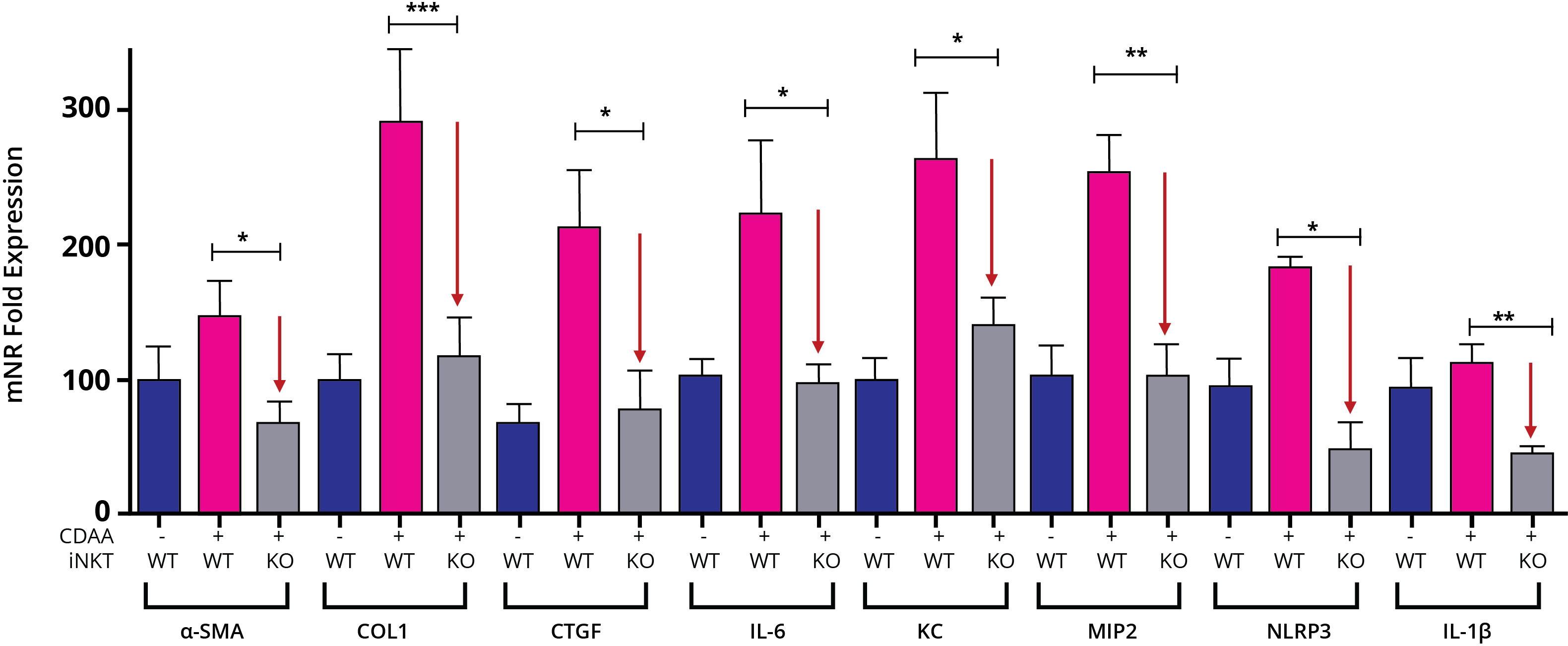
iNKT cell deficiency downregulates expression of key pro-inflammatory and pro-fibrotic genes in a NASH model of liver fibrosis.
Pro-inflammatory and pro-fibrotic genes are elevated in wild type animals fed a choline deficient diet (CDAA, red bars) compared to animals fed a control diet (blue bars); however, CDAA diet-induced increase in the expression of these genes is significantly diminished in animals deficient in iNKT cells (grey bars).
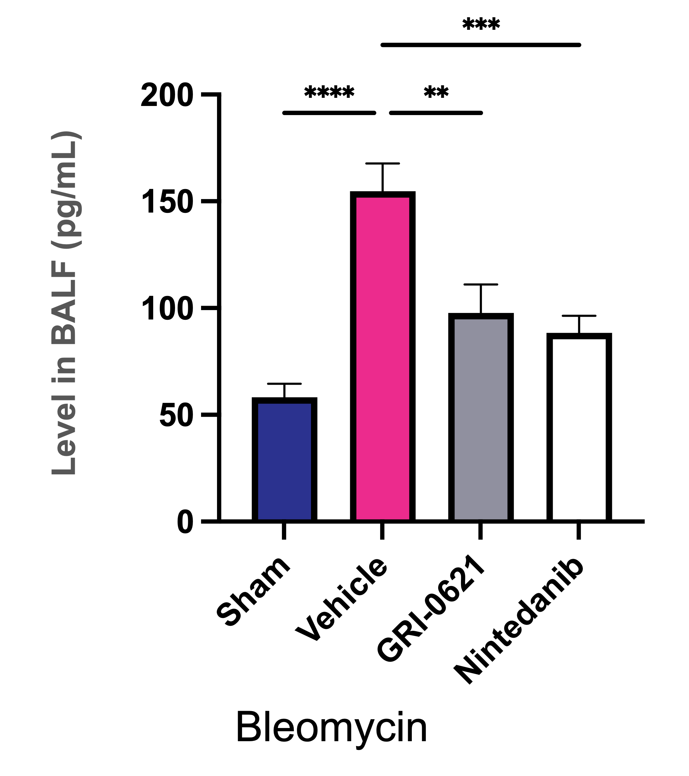
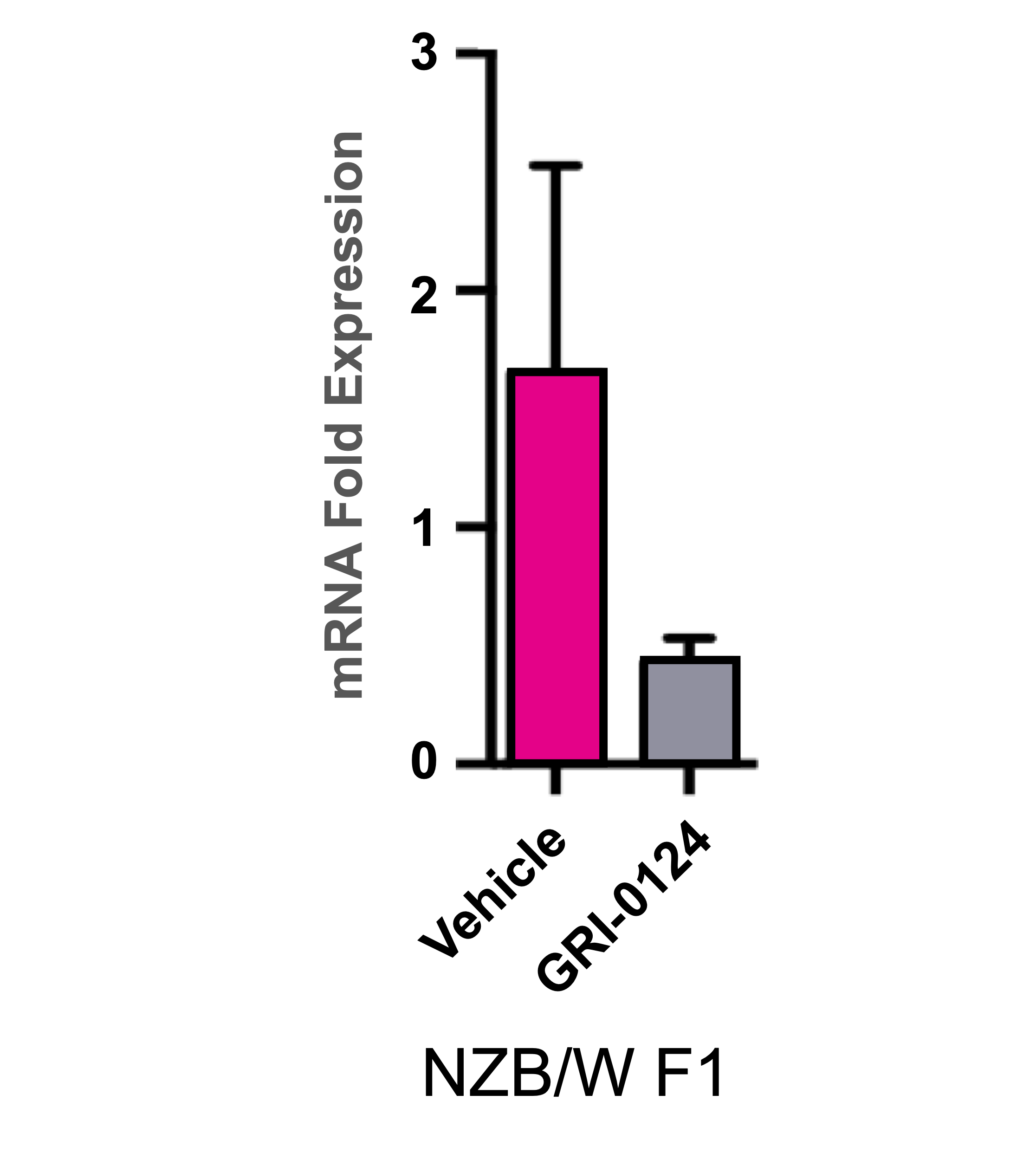
Modulation of NKT Activity Results in Inhibition of TGF-β in Multiple Fibrosis Models
TGF-β is a Central Mediator of Fibrogenesis in Pulmonary, Hepatic and Renal Fibrosis
Pulmonary Fibrosis
Hepatic Fibrosis
Renal Fibrosis
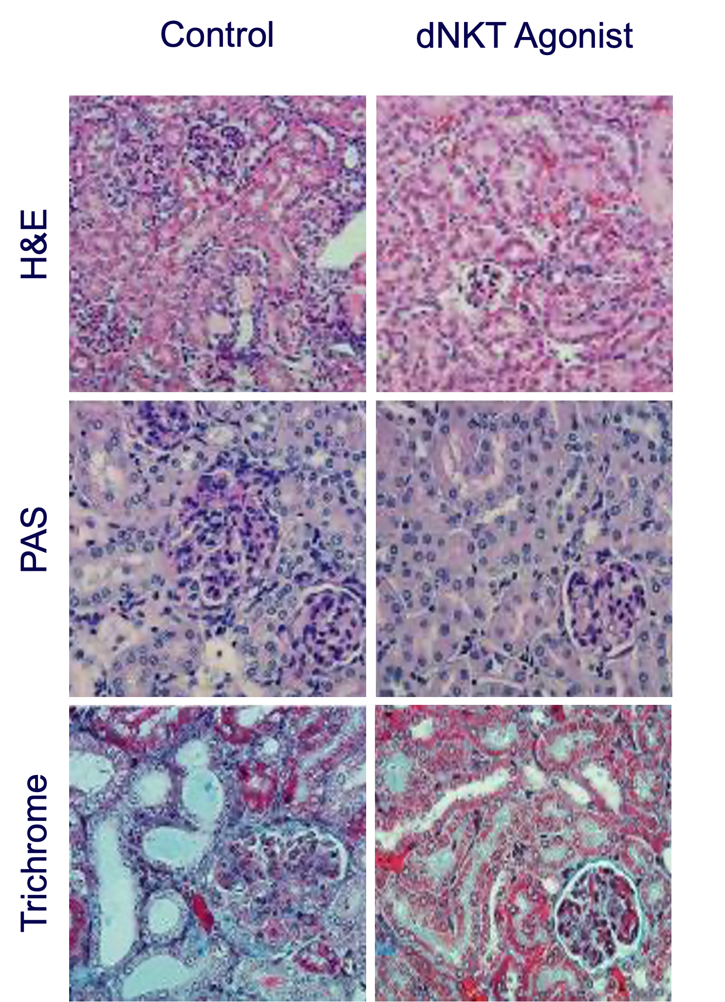
Inhibition of iNKT cell activity reduces the production of TGF-𝛽, a key pro-fibrotic cytokine, in models of pulmonary, hepatic, and kidney fibrosis.
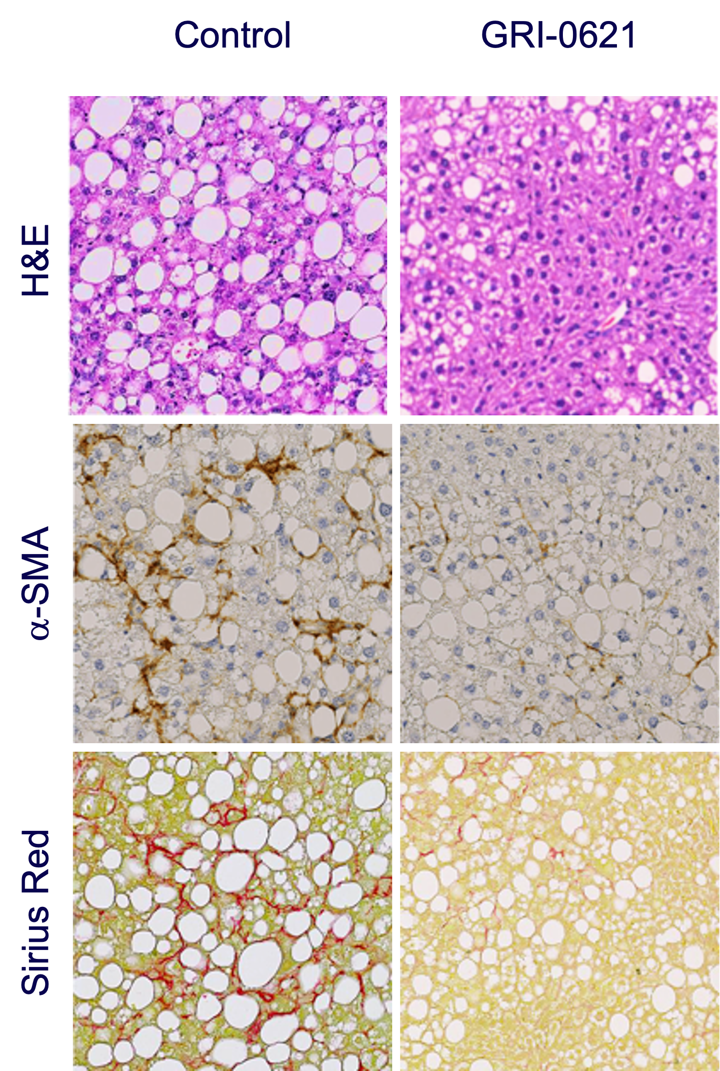
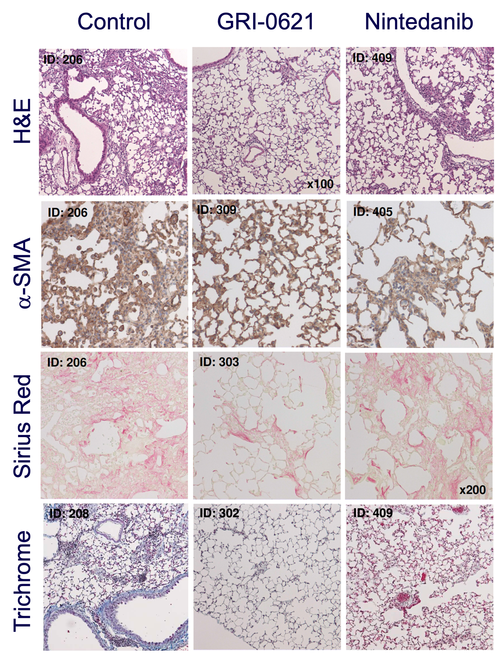
Observed Reduction of Inflammation and Fibrosis in Fibrosis Models
Hepatic Fibrosis
Pulmonary Fibrosis
Renal Fibrosis
